Benchtop TEM microscope LVEM5
TEM, STEM, SEM the compact 3 -in- 1
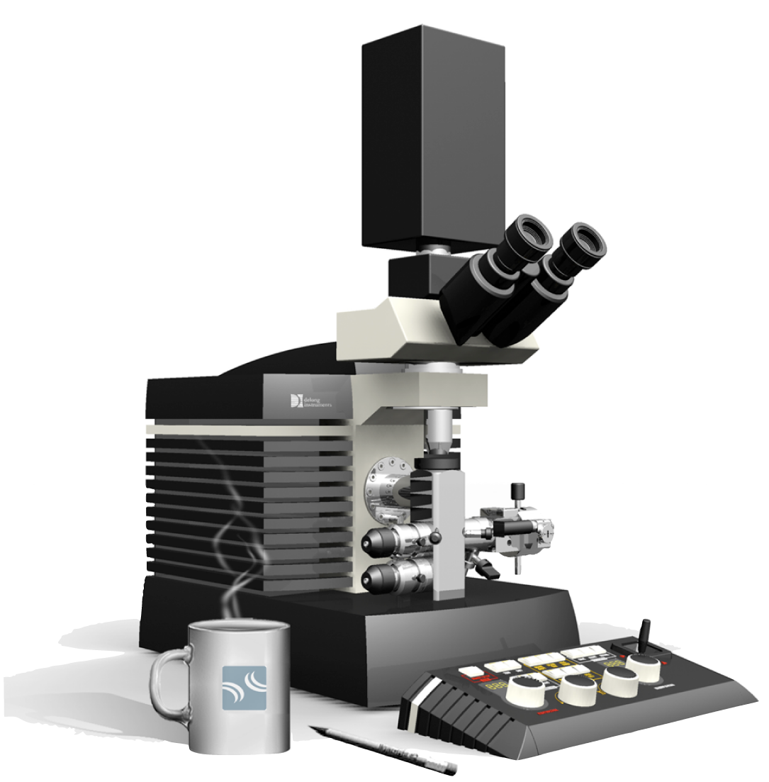
The first benchtop electron microscope that combines TEM, SEM & STEM imaging modes
The smallest TEM in the world…
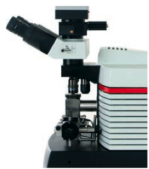
The LVEM5 is a compact benchtop instrument that combines high resolution imaging with the small footprint of an optical microscope. It consists of four separate parts; the microscope, the electronics unit, the vacuum system, and the PC. Small footprint, no need for a dark room, no cooling water, easy service… all this makes the instrument a multi-purpose personal or in-group electron microscope.
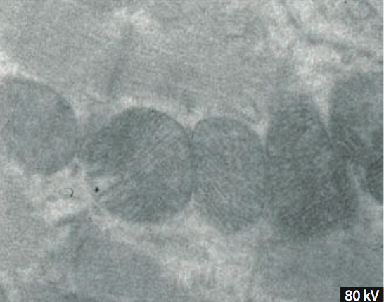
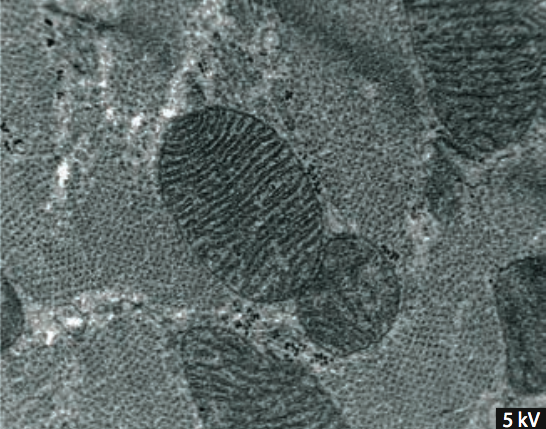
High contrast
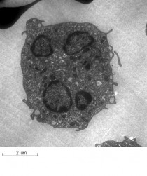
The LVEM5 is a unique investigation tool that allows observation of objects composed of light elements with high contrast without using heavy metal staining and shadowing.
Classical TEM Unstained thin section of rat heart (80 kV) | LVEM 5 Unstained thin section of rat heart (5 kV) |
A wide choice of imaging modes
The LVEM5 is the smallest commercial transmission electron microscope in the world, and features all the standard imaging modes that can be found in conventional TEMs and more. The LVEM5 can work in transmission (TEM – Transmission Electron Microscope) or diffraction (SAED – Selected Area Electron Diffraction) modes as well as in scanning modes (STEM– Scanning Transmission Electron Microscope and SEM – Scanning Electron Microscope avec BSE – Backscattered Electrons) with nanometer spatial resolution.
Components
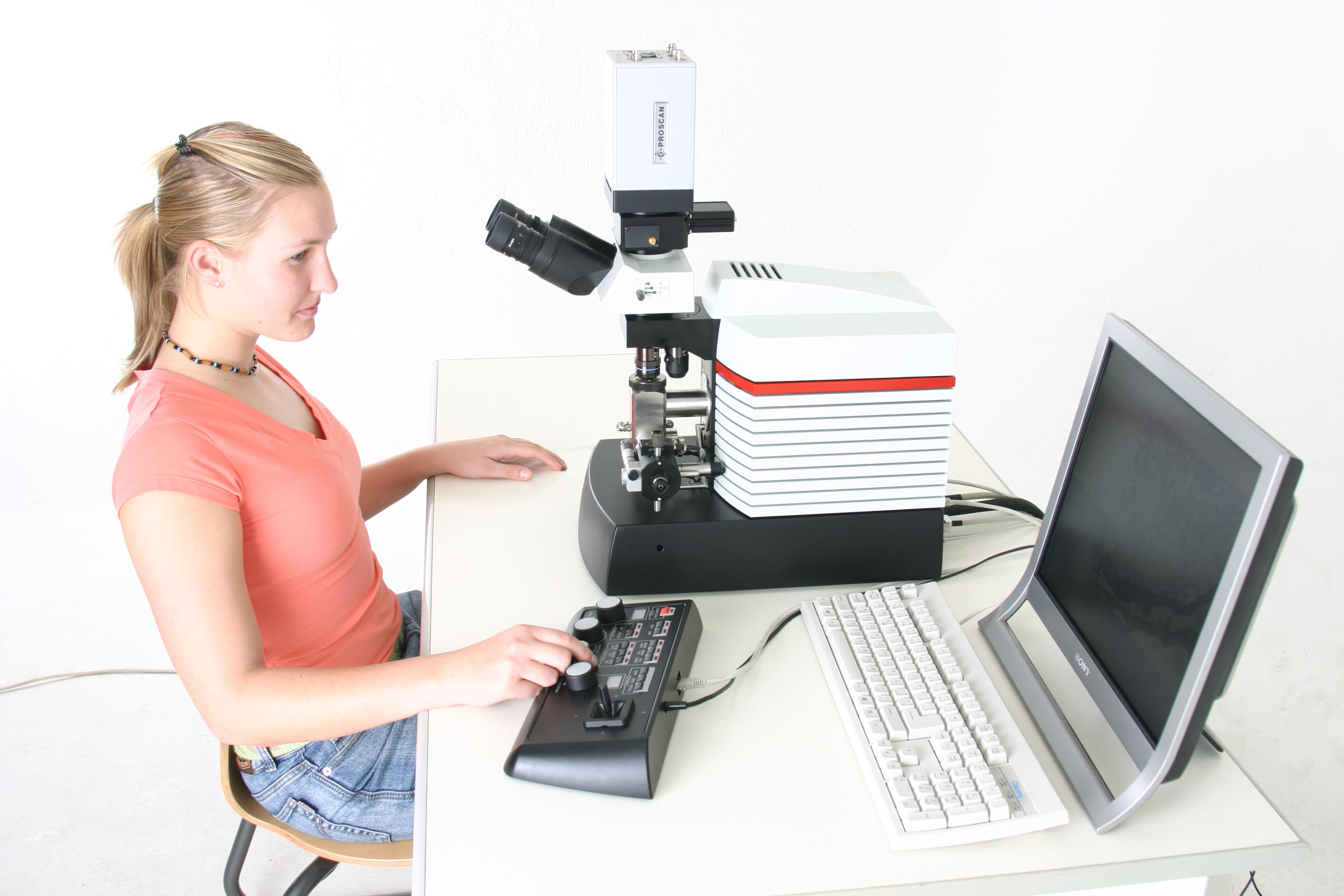
The electron gun uses a Schottky field emitter which provides high brightness and coherence with a lifetime of several thousand hours. The high brightness and small virtual source of the electron gun allows transmission and scanning modes.
Permanent magnet lenses, an electrostatic lens and electrostatic stigmators and defl ectors are used in the electron optics. Permanent magnet lenses are very stable and do not need any cooling.
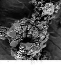
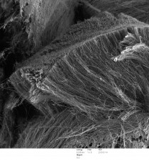
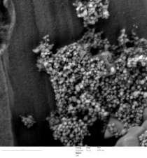
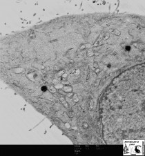
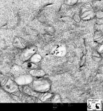
LVEM5 SEM mode gallery
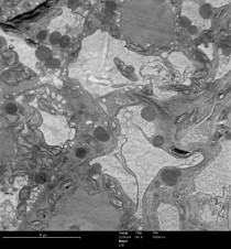
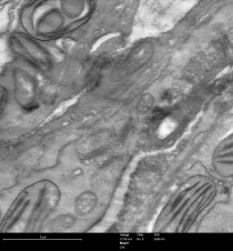
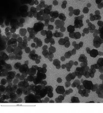
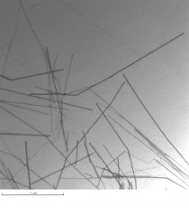
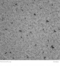
LVEM5 TEM mode gallery
Benchtop TEM microscope LVEM5
Accelerating voltage (nominal) | 5kV | |||
Specimen | standard diameter 3.05 mm grids | |||
time for sample exchange | approx. 3 min | |||
. | ||||
Imaging modes | ||||
TEM | ||||
resolving power | 2.5 nm | |||
total magnification* | 1 500 to 202 000 | |||
*depending on the camera ship | ||||
. | ||||
STEM | ||||
resolving power | 2.0 nm | |||
minimum magnification | 6,000 (25 × 25 µm) | |||
. | ||||
SEM | ||||
resolving power | 2.0 nm | |||
minimum magnification | 800 (200 × 200 µm) | |||
. | ||||
ED (electron diffraction) | ||||
minimum probe size | 100 nm | |||
diffraction lens | magnification 3.5 | |||
. | ||||
Electron optics | ||||
Condenser lens | spermanent magnet | |||
focal length* | 4.30 mm | |||
the smallest illumination area | 100 nm | |||
condensor apertures | 50, 30 micron | |||
*calculated for 5 kV | ||||
Objective lens | permanent magnet | |||
focal length* | 1.26 mm | |||
CS (spherical aberration coefficient) | 0.64 mm | |||
CC (chromatic aberration coefficient) | 0.89 mm | |||
theoretical resolution | 1.1 nm | |||
theoretical aperture angle | 10-2 rad | |||
objective aperture | 50, 30 µm | |||
*calculated for 5 kV | ||||
Projection lens | electrostatic | |||
magnification on the YAG screen | 36–470 | |||
Electron Gun | ||||
SE cathode ZrO/W[100] | ||||
current density | 0.2 mA sr-1 | |||
lifetime | > 2,000 hours | |||
. | ||||
Vacuum | ||||
Airlock system | ||||
diaphragmal pump | 10-5 mbar | |||
. | ||||
Object space | ||||
ion getter pump (10 L.sec-1) | 10-8 mbar | |||
. | ||||
Electron gun | ||||
ion getter pump (7 L.sec-1) | 10-9 mbar | |||
. | ||||
Light optics | ||||
objective Olympus M 40x | ON 0.90 | |||
objective Olympus M 4x | ON 0.13 | |||
binocular M 10x | ||||
TEM image capture | ||||
camera | Retiga 4000R CCD | |||
resolution | 2048 × 2048 pixels | |||
digitalization | 12 bits | |||
pixels size | 7.4 × 7.4 µm | |||
cooling | optional Peltier cooling available | |||
SCAN modes image capture | ||||
monitor | 512 × 512 pixels | |||
saiving image | jusqu’à 2048 × 2048 pixels | |||
digitalization | 8 bits | |||
. | ||||
Weight and dimensions | ||||
Electron and light optic system | ||||
weight | 25 kg | |||
dimensions (WxDxH) without camera | 290 × 450 × 430/480 mm | |||
. | ||||
Airlock pumping system Pfeiffer Vacuum TSH 071E | ||||
weight | 15 kg | |||
dimensions (WxDxH) | 300 × 300 × 340 mm | |||
. | ||||
Control electronics | ||||
weight | 19 kg | |||
dimensions (WxDxH) | 470 × 270 × 290 mm | |||
. | ||||
Electric consumption | ||||
Control electronics in stand by (ion getter pump only) | 20 VA | |||
Control electronics | 160 VA | |||
Including airlock pumping system | 300 VA | |||
Camera | 24 VA | |||
PC and monitors approx. | 450 VA | |||
No cooling water for the microscope operation is required |
Benchtop TEM microscope LVEM5
LVEM5 in Nano-Science Education…
The LVEM5 tool will allow you to introduce students to all areas of nanoscale. Real hands-on experience with three different types of Nano-imaging techniques commonly used in industry will certainly give your students a competitive edge upon entry into the workforce…
Nanomedicine, how the LVEM5 helps ?…
With the LVEM5 you will be able to resolve the sizes of your nanoparticles with improved contrast to understand the quality of your synthesis on a number of levels. You will be able to discern particle size, quantity and distribution. This can all be easily accomplished in minutes on the LVEM5, right in your own lab. You get all this for a fraction of the price of a conventional electron microscope…
Polymer Science…
The LVEM has proven to be particularly useful for the high contrast imaging of a wide variety of polymers, organic molecular thin films, and biological materials. We have obtained images on a variety of systems including polyethylene single crystals, pentacene and TIPS-pentacene thin films, block copolymers, and poly(3,4-ethylene dioxythiophene…
…
SAMPLE PREPARATION GUIDE
Select the characteristics of your sample among the criteria (Organization of material, physical state…) then click on the button “confirm your choices”. Your choices will orient you towards one or several techniques, click on to see details (advantages, drawbacks…). To deepen your search, refer to the “pedagogical guide”.